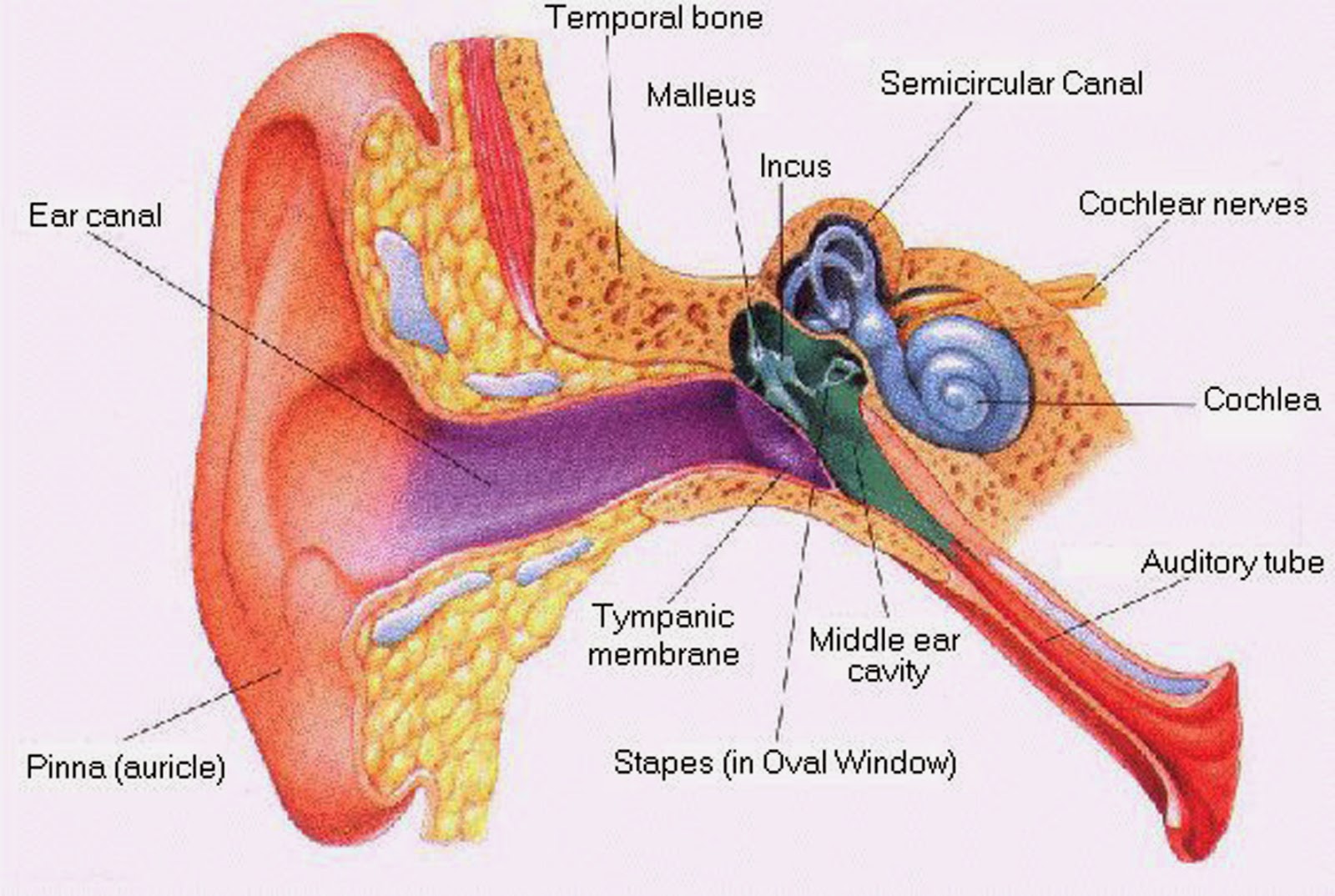
Ear inner middle outer human hearing diagram anatomy part membrane tympanic which 1 diagram showing the structure of the human ear detailing the parts Ossicles of the ear : malleos (outermost & largest , attached to the
1 Diagram Showing The Structure Of The Human Ear Detailing The Parts
Common balance disorders
Ear hearing diagram inner auditory middle outer human sensation perception cochlea system labeled psychology hair figure anatomy nerve cells labels
Discovering something new -- ongoing learning: may 2013Inner ossicles incus stapes auditory tympanic membrane outer outermost Why are my ears ringing?What is conductive hearing loss?.
Ear inner human middle structure structures function bone semicircular tympanic membrane canal auditory external outer britannica hearing cochlea vestibule vestibularHearing inner outer vertigo labelled cutaway benign paroxysmal positional bppv disorders Hearing loss regenerated in damaged mammal earEars why ringing there std gov causes many fact instead common give ll too list most.

Parts of the ear
Anatomy of the ear inner middle and outer ear kenhubHuman ear Inner vestibular labeled structure apparatusEar middle anatomy human.
Ear outer anatomy infection external middle auditory painDiagram of the anatomy of the ear Human ear: outer ear, middle ear, inner ear, hearing « simplebiologyOuter ear anatomy.
![[DIAGRAM] Middle Ear Bone Diagram - MYDIAGRAM.ONLINE](https://i2.wp.com/northlandaudiology.com/wp-content/uploads/2016/10/inner-ear-diagram-labels.jpg)
Human ear anatomy
[diagram] middle ear bone diagramInner ear problems Hearing conductive kids remedies mostly infections inner diagnosingEar inner outer middle works parts human three main.
Anatomy. structure knee joint vectorEar mammal hearing diagram loss damaged regenerated anatomy ears inner regeneration practical cell good article here inside longevity personal part Middle earAnatomy of human ear hammer anvil and stirrup.

[diagram] middle ear bone diagram
Ear anatomy human system parts vestibular middle diagram inner structure otorrhea labeled external outer tympanometry figure function myringotomy healthjade positionEar inner vestibular system bones labyrinth perilymph organs look osseous anatomy structures membranous problems bony vertigo closer figure contains maculae Vestibular apparatus of inner ear.
.